
Advanced endodontics

Root Canal Treatment Under the Microscope: You Can Only Treat What You Can See

The dental operating microscope magnifies the working area in root canal treatment by four to twenty-five times and lights it with coaxial LED illumination; it is one of the most important technological advances in endodontics. Extra canal orifices, calcifications, micro-cracks and fragments of broken instruments that the naked eye cannot pick out all become visible under the microscope.

At our clinic, endodontics under the microscope — the advanced practice of what the international literature calls the endodontist — is carried out by Dr G. Cansu Öztürk, endodontic specialist. Specialist training and the microscope together are the two factors that decide how predictable treatment is, particularly in complex and retreatment cases.
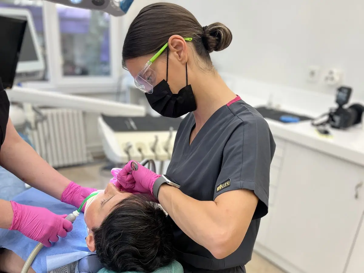
Root canal treatment under the microscope — dental operating microscope, GC Clinic Nişantaşı
Why it matters

The limit of the naked eye should not be the limit of the treatment.

The root canal system is not a simple tube: it has lateral branches, curves, extra canals and variable anatomy. Upper molars frequently have a second mesiobuccal canal, known as MB2, which is easily missed with the naked eye or at low magnification. Every canal left untreated is among the most common causes of later infection and of the need for root canal retreatment.

The microscope reduces that risk at source: canal orifices are found by looking at them directly, the cleaned area is confirmed by sight rather than by assumption, and the limits of the filling are checked. It also means less healthy tissue is sacrificed — because the dentist can see where to open, the access cavity stays small. This is what our conservative approach means in endodontics.

Indications

The cases where a microscope makes the difference.

Hidden and extra canals (MB2)

Atypical anatomy — the second mesiobuccal canal in upper molars, a second canal in lower incisors — is found under the microscope and included in the treatment.

Calcified (narrowed) canals

Canal orifices blocked by calcification from age or injury are opened safely under magnification, with no blind searching.

Root canal retreatment

Removing the old filling material completely, finding the canals that were missed and reshaping them are all done under microscope control.

Removing broken instruments

Fragments of file left in the canal by a previous treatment are removed with ultrasonic tips under microscope vision.

Repairing perforations

Unintended openings in the root wall are repaired with bioceramic materials, with the damaged area in direct view.

Diagnosing a cracked tooth

Micro-cracks are examined under magnification and whether the tooth can be saved is decided on the evidence — no treatment is proposed for a tooth that cannot be saved.

The process

How does treatment under the microscope proceed?

1

Imaging

The root anatomy is mapped by examination and, where the case calls for it, a CBCT (three-dimensional) scan.

2

Isolation

The tooth is isolated from saliva with a rubber dam, creating a sterile working field.

3

Treatment under the microscope

The canals are located, cleaned, shaped and filled under magnification and coaxial light.

4

Checking and restoration

The limits of the filling are confirmed, and the treatment is completed by planning the restoration on top — a filling, an onlay or a crown.

Comparison

Is a microscope different from magnifying loupes?

Dental operating microscope

Stepped magnification from 4x to 25x · coaxial LED light (shadow-free vision) · a stable, shake-free image · the ability to document the case · the standard tool for MB2, perforation and broken-instrument cases.

Magnifying loupes

Usually a fixed magnification of 2.5x to 4.5x · a head-mounted light · useful for routine work, but they do not reach the level of vision a microscope gives on fine anatomical detail.

Success and longevity

What determines success in root canal treatment?

The long-term success of root canal treatment rests on three things: finding and cleaning every canal, filling the canal system so that it does not leak, and restoring the top of the tooth properly. In a large share of failed cases the problem is a canal missed at the first attempt or an area left incompletely cleaned — in other words, a problem of vision.

The microscope strengthens the first of those three directly: canal orifices are found by sight, not by assumption. That is why the endodontic literature has made magnification and illumination the standard for advanced cases. At our clinic the last link is completed in-house too: the restoration on top is planned under the same roof, in our in-house laboratory.

Who it is for

Assessment under the microscope matters most in these situations.

People having root canal treatment on a molar

Molars have three or four canals, sometimes more; they are the group of teeth most likely to have an extra canal. Getting the first treatment right prevents the need for retreatment altogether.

People whose root canal treatment has failed

For teeth where the pain does not settle, an abscess keeps returning or an X-ray shows a lesion that is not healing, retreatment is planned under the microscope.

People who want to save a tooth rather than have it out

Many teeth that have been written off as “this one has to come out” turn out to be savable when assessed under the microscope. It is not too late for a second opinion.

Location

Specialist endodontics in Nişantaşı.

Our clinic is in Teşvikiye, Nişantaşı, within walking distance of Osmanbey metro station and easily reached from Şişli, Maçka and the surrounding districts. Root canal treatment under the microscope is carried out by a specialist at only a small number of clinics across Istanbul, and some of our patients travel from outside the city for root canal treatment and retreatment — you are welcome to send us any existing X-rays on WhatsApp before your assessment appointment.

Frequently asked

About root canal treatment under the microscope.

Is a microscope necessary in every case?

Routine front-tooth canals can be treated successfully without magnification; but for molars, calcified canals, retreatment cases and complications, a microscope markedly improves how predictable the treatment is. At our clinic, endodontic cases are assessed with the microscope.

Does it make treatment take longer?

Appointment times are similar; the microscope shortens the searching and speeds up the checking. Complex cases may need more than one appointment — that comes from the nature of the case, not from the microscope.

Is treatment under the microscope painful?

No; the treatment is carried out under local anaesthetic. The microscope only magnifies what the dentist sees and does not change the experience for you — if anything, less tissue is removed, so healing is usually more comfortable.

My root canal treatment failed at another clinic — can the tooth be saved?

In many cases yes: a missed canal, an inadequate filling or reinfection can be put right with retreatment under the microscope. The situation is first clarified with a CBCT scan, and where the tooth cannot be saved you will be told so honestly.

Is root canal treatment guaranteed?

Our root canal retreatment work carries a two-year written guarantee, and the terms are given to you in writing along with your treatment plan.

Who carries out the treatment?

Dr G. Cansu Öztürk, endodontic specialist — specialist training in the Department of Endodontics at Marmara University, member of the ESE, AAE and TED.

Contact

Message us with a question or to book.

You are welcome to a free consultation for an assessment under the microscope or a second opinion.

Message us on WhatsApp